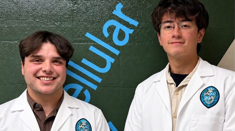
New Orleans – While volunteering in the cadaver lab at Tulane University’s School of Medicine, sophomore students Dominic Roy and Cooper Bruce stumbled upon a previously undocumented muscle attachment in the hip. Their finding, confirmed by professor of neurosurgery R. Shane Tubbs, Ph.D., has been published in the peer‑review journal Clinical Anatomy and may influence future hip‑surgery techniques.
How the discovery happened
Roy and Bruce were dissecting several hip specimens to study the relationship between the iliacus and the psoas major muscles. When Roy was looking for the iliocapsularis, a muscle that lies laterally in the hip, he noticed a similar‑looking structure on the opposite side of the iliac fascia, near the inguinal region. “I called Dr. Tubbs over and asked, ‘What is this?’” Roy recalled. Tubbs, who also serves as co‑editor‑in‑chief of Gray’s Anatomy, admitted he had never seen the structure before.
Research and verification
The students conducted an extensive literature review and consulted textbooks, but found no prior description of the muscle. Their team, which included physicians from overseas and other medical students, photographed the specimen and documented the anatomy. Tubbs described the discovery as “serendipity,” noting that the muscle runs deep in the pelvis and extends to the front of the hip joint.
Potential clinical impact
Although the exact function of the new attachment remains unknown, early clues suggest it may help stabilize the psoas major, a large muscle involved in hip flexion. Tubbs explained that future research will focus on whether magnetic resonance imaging (MRI) can reliably detect the attachment and whether its presence correlates with patient symptoms.
If the muscle proves to be a source of pain or weakness, surgeons could consider targeted physical‑therapy regimens or, in some cases, injections to reduce overactivity. “Surgeons are replacing hips and doing hip surgery on a daily basis, so if they see the muscle, potentially they could correlate the symptoms of the patient and whether cutting the muscle would help,” Tubbs said.
Broader significance
The discovery underscores the value of hands‑on cadaver work in medical education. Tubbs warned that synthetic models, while useful, cannot replicate the natural variation found in real human bodies. “When you look beneath the layers of the skin, human beings have lots of variation,” he noted, adding that no two individuals are built the same inside or out.
Interestingly, the same muscle attachment was observed in gorilla specimens, hinting at an evolutionary link. Bruce speculated that lifestyle factors such as frequent soccer or rock‑climbing might influence the development of the attachment in some individuals.
Next steps for the researchers
Roy and Bruce, now seniors, plan to continue their studies and hope the attachment will eventually be added to anatomical textbooks. Roy intends to pursue a master’s program in anatomy, while both students see the experience as a catalyst for future medical careers.
The finding reminds the medical community that anatomical knowledge is still evolving, even in the 21st century. As Tubbs concluded, “Many people think we’ve known every part of the human body for hundreds of years, but in actuality, people are still discovering things about human anatomy.”
Original reporting: Fox News (HLL/CB) — read the source article.