
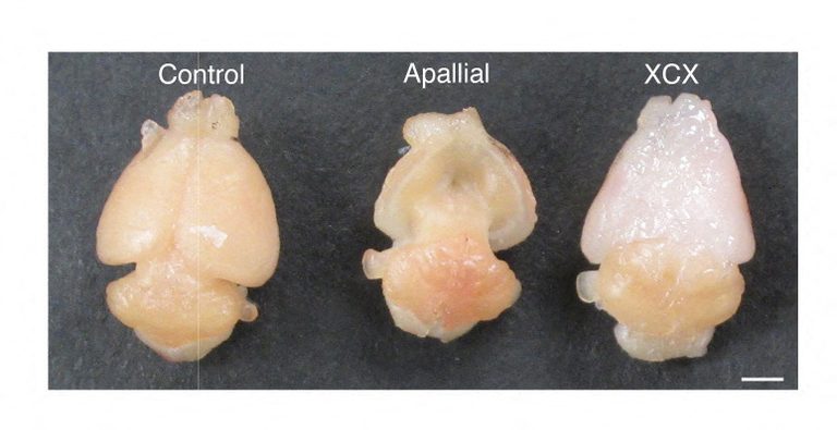
In a breakthrough that could speed research into devastating neurological conditions, scientists have successfully transplanted laboratory‑grown human brain tissue into specially engineered mice. The animals, dubbed “xenocortical” mice, carry a larger volume of human cortical tissue that integrates with the mouse nervous system.
How the model was built
The team, led by Stanford University neuroscientist Dr. Sergiu Pasca, first created three‑dimensional cortical organoids—mini‑brain structures that mimic key cell types of the human cerebral cortex. These organoids were derived from reprogrammed skin or blood cells, which were turned into stem cells capable of becoming any cell type.
To make space for the human tissue, the researchers used a genetic strategy that blocks development of most mouse cortical and hippocampal cells. The resulting mice lack the outermost layer of the brain, allowing the human organoids to be transplanted shortly after birth and to grow extensively.
Human tissue thrives in the mouse brain
Once implanted, the human grafts generated a broad diversity of cortical cell types, including the rare von Economo neurons, which are vulnerable in certain dementias. The grafts formed functional connections throughout the mouse nervous system, demonstrating that the human tissue can develop and integrate in a living animal.
Dr. Pasca emphasized that these mice are not “mini‑brains” or “humanized mice.” Rather, they retain a mouse nervous system while housing a substantial amount of human cortical tissue that develops, integrates, and forms connections within the host.
Potential for disease research
The new model offers an experimental window into human brain development and disease. Researchers can now study how disease‑associated genetic changes affect neural development, circuitry, and response to potential treatments—tasks that are ethically impossible with living human brain tissue.
In a first application, the team exposed the xenocortical mice to low‑oxygen conditions that mimic birth‑related oxygen deprivation. The human cortical cells showed substantial injury and the mice displayed gait and motor coordination deficits, while ordinary lab mice were unaffected. This suggests the model can help investigate conditions such as cerebral palsy, epilepsy and autism that often arise from perinatal oxygen loss.
Ethical safeguards
The researchers stressed adherence to two core ethical principles. First, animal welfare: the scientific question must justify animal use, suffering must be minimized, and alternatives must be exhausted. Second, the introduction of increasingly complex human neural tissue into an animal raises questions about emergent properties that may require further ethical review.
Dr. Pasca noted that the cost of not pursuing this work is high, given that neurological and psychiatric disorders affect nearly one in five people, yet effective treatments remain limited.
What this means for the future
While the xenocortical mice do not replicate the full complexity of the human brain, they provide a valuable platform for studying human neural cell types, developmental processes and disease mechanisms that are otherwise inaccessible. The approach could accelerate the discovery of therapies for autism, epilepsy, schizophrenia, cerebral palsy and related conditions.
Original reporting: Appleton, WI News Feed (HLL/CB) — read the source article.